Major Organs in the Circulatory System
Heart
|
|
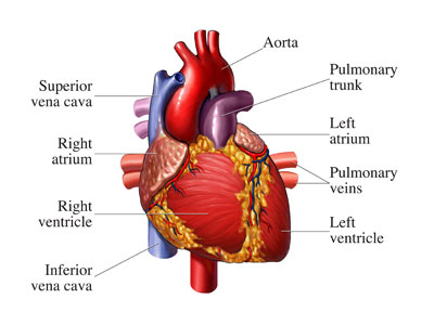
The heart is a hollow, muscular pump located in the left-center of the chest that has two sides that work in tandem to pump a total of 5L or 9 pints of blood around the body. The heart's cardiac muscles cannot receive oxygen from the blood they pump, so there are two coronary arteries that split off from the aorta to supply the chambers with oxygen. This is to make sure that the heart's muscles can continue contracting without tiring or needing a break.
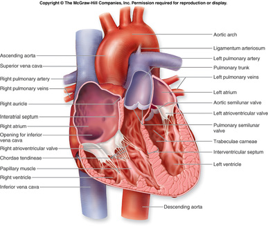
The right side of the heart receives oxygen-poor blood from the body and pumps it to the lungs for exchange, while the left side of the heart receives oxygen-rich blood from the lungs and pumps it around the body. The heart is composed of mainly two ventricles and two atria (one on each side). The right atrium receives blood from the superior and inferior vena cava. The left atrium is supplied by a pair of pulmonary veins (one from each lung). The left ventricle exports oxygen-rich blood along the aorta throughout the body. The right ventricle carries oxygen-poor blood through the pulmonary arteries to both lungs.
The heart has a natural pacemaker that's situated to te left of the heart that tells the heart when to beat. When you're excercising, the pacemaker causes your heart to beat faster so all of the hard-working muscles have enough oxygen, but when you're resting, it tells your heart only to beat quick enough to sustain life. Normally your heart beats about 70 time per second when you're resting.
stages of heartbeat
= atrial systole = ths atria contract together to pump blood into the ventricles while opening the valves between the two chambers
= ventricular systole = ventricles contract to force blood out of the heart and at the same time the valves between the atria and ventricles close
= diastole = the cardiac muscles relax to let blood flow into the atria and the valves for exiting the ventricles
Blood Vessels - Capillaries, Arteries, and Veins

|
|
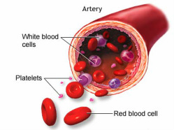
Blood vessels transport all of the vital substances to keep cells alive as well as red and white blood cells and platelets. Pumped by the heart, oxygen-rich blood travels to all the parts of the body so oxygen-rich blood cells can diffuse oxygen into the body cells, and so body cells can diffuse carbon dioxide into the red blood cells so it can be breathed out before it poisons the body. There are three types of blood vessels: arteries, veins, and capillaries.
Arteries are thicker and they carry oxygen-rich blood from the heart to the body. They're tougher so they can withstand the high-pressure pump of blood. Arteries also have a slick inner lining to reduce friction and let blood flow smoothly. Arteries have a protective outer coat, a thick layer of muscle and elastic fibers, elastic and connective tissue, and a slick inner lining.
Veins are thinner and less muscular because they don't have to withstand the high-pressure pump of blood, but since veins aren't very strong many of them have valves to make sure that blood doesn't flow backwards. They bring blood back to th eheart so it can be refueled with oxygen and can dump the waste carbon dioxide. Veins have an outer layer, a thin muscle layer, valve(s), and a slick inner lining to let blood flow smoothly.
Capillaries are very small, no wider than one blood cell, which means that they're about 0.0008mm or 0.0003in in width! They connect the arteries and veins and form networks called capillaty beds. Capillaries are 98% of the total length of all blood vessels and there are more than 40 billion capillaries in the body! Since capillaries are so thin, they're very leaky and allow oxygen and food to easily pass through. They come from arterioles, which are small arteries, and will eventualy merge into venules, which are small veins, and further into veins to travel back to the heart.
stages of blood vessels
= artery -> arteriole -> capillary -> venule -> vein -> REPEAT
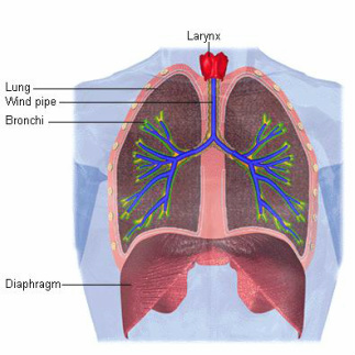
Lungs (Bronchi, Bronchioles, and Alveoli)
|
|
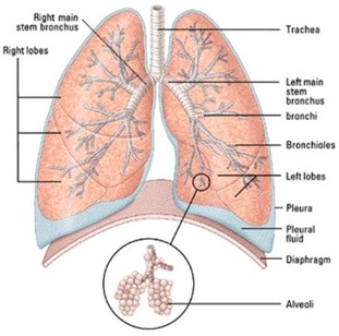
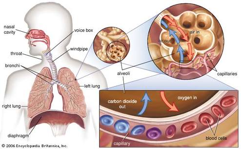
The lungs contain the bronchi, bronchioles, and alveoli. It's protected by the ribcage along with many other organs. The bronchi in each lung continue to divide until they are small enough to turn into bronchioles. The smallest bronchioles end in microscopic airbags called alveoli. Alveoli are the organs that exchange oxygen and carbon dioxide between the air and the blood. Alveoli are very efficent because they have a network of capillaries that cover them and the fact that there's 300 million alveoli in the lungs. This provides a massive area for efficently transferring oxygen and carbon dioxide through the alveolus walls as quickly as possible.
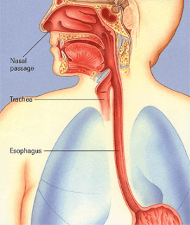
Trachea
|
|
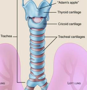
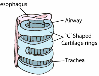
The trachea, or windpipe, connects the lungs to the mouth and itself is the piece that connects the two lungs together. The trachea has horizontal "C"-shaped rings around the windpipe that act as reinforcements so the trachea doesn't collapse in on itself when air is breathed in. The inside is lined with cilia and mucus that continue the jobs of removing airborne germs and dirt. At the lowest end of the trachea, it branches off into two bronchi (one for each lung).
Diaphragm
|
|
The diaphragm is located underneath the lungs and is the muscle that causes the lungs to breathe air in and out. Muscles in the diaphragm relax and are pushed upwards at the same time when we exhale, and are contracted downwards away from the lungs to inhale air into the empty space. When irritated, it suddenly contracts downwards which sucks in air. This causes the vocal cords in the larynx to snap shut and produce a "hic" sound. This is how hiccups are caused.
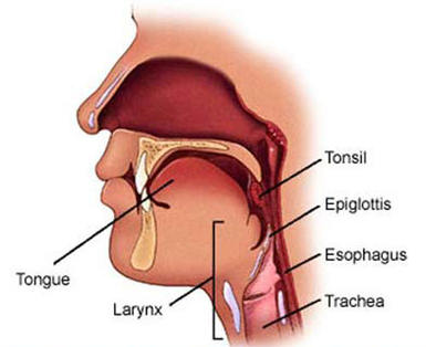
Esophagus (Larynx and Epiglottis)
|
|
The esophagus is commonly known as the throat, and it is the tube where food and air are obtained. The larynx is the part of the esophagus where air is taken in. You are able to see the larynx at the front of your throat. The larynx, or voicebox, connects the trachea to the mouth. The larynx, also known as the "Adam's Apple", is made of several plates of cartilage. The epiglottis is a hinged flap that decides which pipe something goes down. The epiglottis closes for the pharynx (food) and opens for the larynx (air). Without the epiglottis things would always be going down the wrong pipe.
Kidneys
|
|
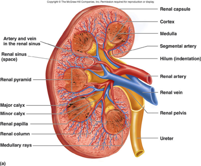
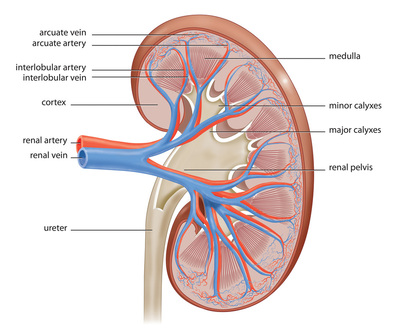
The kidneys filter blood to remove wastes from the body and release them as urine. The darker-coloured outer region of the kidney contains one million filtration units called nephrons that filter the blood containing both wastes and useful substances into a tubular nephron. The renal vein carries the "cleaned" blood from the kidney back to the heart, where as the renal artery carries oxygen-rich blood to the kidney. As blood passes throught the nephron, useful substances like glucose diffuse back into the bloodstream while unwanted wastes, water, and salts form urine and driblle down into the kidney's pelvis and then throught the ureter into the bladder.
Liver
|
|
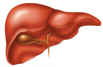
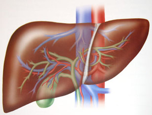
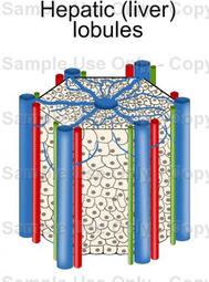
Liver cells produce a yellow-green liquid called bile that is mainly made of water, but also bile salts that help digest fats and wastes . Bile is stored in the gallbladder and lines the small intestine. The liver diffuses vital nutrients into the bloodsrteam to ensure that they're stored or dispatched where it's needed and has white blood cells thta help remove bacteria and debris from the blood. The liver is an unusal organ because it has two blood supplies instead of one. The hepatic portal vein carries oxygen-poor, nutrient-rich blood from the small intestine that's ready for processing by the liver cells. The hepatic artery delivers oxygen-rich blood to the liver cells. The liver cells are organized in processing units called liver lobules. The combined activities of the liver cells release heat that help keeps the body warm.
The liver is a very important organ because it carries out more than 500 functions! A few of the liver's jobs are:
= maintains glucose (body's fuel) level by storing and releasing it when needed
= stores and packages fat for transport
= uses amino acids to make proteins
= processes amino acids to make urea
= makes bile (used to digest fat that includes substance that were once in red blood cells)
= breaks down drugs and poisons (eg. alchohol)
= removes hormones from blood to stop them from working
Liver Lobules
|
|
Liver lobules are the liver's processing plants and each of them is no bigger than a sesame seed. Lobules are vertical sheets of cells that radiate from the central vein which processes the blood that is to be returned to the heart. At the corners of each lobule, there's a hepatic vein that delivers nutrient-rich blood, and a hepatic artery that delivers oxygen-rich blood. The two rich bloods are mixed and processed by liver cells as they travel along wide capillaries towards the central vein.
